Tag Archives: orthopaedic implant manufacturer

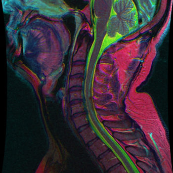
Time to seriously consider focused mris for injured spines

Is it sufficient to only examine the injured part of the spine via focused magnetic resonance imaging (FMRI) or will that lead to missed problems?

In patients who sustain blunt trauma, CT is the initial screening modality of choice in most emergency departments in the United States to look for injury to various parts of the body—including the spine. If a fracture is seen in the thoracic or lumbar spine on CT, sometimes an MRI of the injured segment of the spine is useful to provide additional information about the injury to help physicians decide on a treatment plan.”

“The problem is that MRI is a relatively expensive and time-consuming test compared to CT, so every time we were ordering MRIs of the entire spine in this scenario, it seemed somewhat excessive. Since we are mostly interested in looking at the part of the spine that we already know is injured (based on the CT), we asked ourselves if just imaging the injured portion of the spine by MRI (a focused MRI) would be adequate to provide the information we need to decide on a treatment plan. The concern with this approach is whether we would miss injuries by not looking at the entire spine by MRI.”

To answer the question, the investigators reviewed records for all adult trauma patients who presented to Mass General’s emergency department between 2008 and 2016 with one or more fractures of the thoracic and/or lumbar spine—as identified using computed tomography (CT) and who underwent MRI of the entire thoracic and lumbar spine within 10 days.

The most important result of the test, said Dr. Karim, was “that performing a focused MRI of only the injured portion of the spine would lead physicians to missing some sort of injury in another portion of the spine about 15% of the time, but none of these ‘missed injuries’ would lead to any change in treatment plan or patient care if they are known.”

“This is a very practical study from our perspective because it affects the day-to-day work of radiologists, emergency room physicians, and spine surgeons. If they see a fracture in the thoracic or lumbar spine on CT that they want to better characterize on MRI, they should be comfortable obtaining just a focused MRI of the injured portion of the spine—saving time, money, and patient discomfort.”

Reference From : https://ryortho.com

Patient relevant outcomes of unicompartmental vs total knee replacement

Between 1 January 1997 and 31 December 2018, via searching Medline, Embase, Cochrane Controlled Register of Trials (CENTRAL), and Clinical Trials.gov, researchers provided a clear and comprehensive summary of the published data on unicompartmental knee replacement (UKA) or total knee replacement (TKA), comparing domains of outcome that have been shown to be important to patients and clinicians in order to enable informed decision making. Findings suggested that TKA and UKA are both viable treatment options for isolated unicompartmental osteoarthritis arthritis. This investigation shows better results for UKA in several outcome domains by directly comparing the two treatments. The risk of revision surgery for TKA, however, was lower. This information should be available to patients when selecting treatment options as part of the shared decision-making process.

Read the Full Article : https://www.bmj.com/content/364/bmj.l352

Reference From : www.mdlinx.com

Type of Knee Ligament Injuries

Our knee is made up of many important structures, any of which can be injured. The most common knee injuries include fractures around the knee, dislocation, and sprains and tears of soft tissues, like ligaments. In many cases, injuries involve more than one structure in the knee.

Anterior Cruciate Ligament (ACL) Injuries

The anterior cruciate ligament is often injured during sports activities. Athletes who participate in high demand sports like soccer, football, and basketball are more likely to injure their anterior cruciate ligaments. Changing direction rapidly or landing from a jump incorrectly can tear the ACL. About half of all injuries to the anterior cruciate ligament occur along with damage to other structures in the knee, such as articular cartilage, meniscus, or other ligaments.

Posterior Cruciate Ligament (PCL) Injuries

The posterior cruciate ligament is often injured from a blow to the front of the knee while the knee is bent. This often occurs in motor vehicle crashes and sports-related contact. Posterior cruciate ligament tears tend to be partial tears with the potential to heal on their own.

Do spine fusion patients with hip arthroplasty risk dislocation?

Anew study of 42,300 patients explores the question of whether patients undergoing lumbar spinal fusion (LSF) before total hip arthroplasty (THA)—versus after—have an increased risk of dislocation and revision.

Study co-author Arthur Malkani, M.D. of the University of Louisville Adult Reconstruction Program, explained to OTWthe rationale for this large study: “There has been a significant increase in the number of patients undergoing primary THA with a history of prior lumbar spine fusion. Lumbar spine fusion is an independent risk factor for dislocation leading to increased risk of revision.”

“The prevalence of hip dislocation following primary THA in patients with prior lumbar spine fusion in the Medicare population is 7.4%. Lumbar spine fusion alters spino-pelvic anatomy leading to loss of the normal pelvic parameters during standing and sitting which could lead to hip impingement and instability.”

In the study, 28,668 patients underwent lumbar spine fusion and then total hip arthroplasty sometime later and 13, 632 had the reverse—total hip arthroplasty followed by lumbar spine fusion sometime after.

Dr. Malkani told OTW, “In our practice we are seeing more patients presenting with both lumbar spine disease and primary hip osteoarthritis. The purpose of this large database study was to determine if hip instability could be decreased if the hip replacement was done first prior to the lumbar fusion in patients with both lumbar spine disease and hip arthritis.”

“The results from this study suggest a significant advantage for patients to undergo THA first if concomitant lumbar spine and hip pathology are likely to require both lumbar spine fusion and THA.”

“There was a 46% increased risk of dislocation in patients with prior LSF compared to the group that had a THA done first followed by a LSF within 1 year, 60% increased risk of dislocation compared to THA first then LSF at 2 years and 106% increased risk compared to THA first and delayed LSF by 5 years. The greater the interval between index THA and proceeding LSF, the lower the risk of dislocation and THA revision.”

“Instability continues to be the leading cause of failure following primary THA leading to revision surgery. We need to identify high risk patients for instability prior to the primary THA. We need to start asking patients if they have any history of lumbar spine disease during the initial visit prior to the THA. In those patients with concomitant lumbar spine disease and hip osteoarthritis, the arthroplasty and spine surgeons need to collaborate to determine if the hip arthroplasty can be approached first with a delay in spine surgery.”

“Additional work is required to determine the ideal functional acetabular cup position in patients with a rigid lumbosacral-pelvic biomechanical relationship and altered pelvic parameters. Surgeons should look at multiple factors in patients undergoing THA with a history of prior lumbar spine fusion to minimize dislocation such as preoperative assessment of pelvic parameters, ideal surgical approach, maximizing head size, restoring offset, satisfactory implant alignment, and an intraoperative assessment of stability.”

Do You Have Lumbar Spinal Stenosis?

Lumbar spinal stenosis (LSS) is a common cause of pain in the lower back and legs. As we get older, our spines go through changes. This is known as spinal degeneration, and it happens to everyone. That being said, even though it happens to everyone, not everyone has negative symptoms that accompany it. When our spines degenerate, the spinal canal may narrow, leading to a condition known as spinal stenosis.

Age-related spinal wear and tear occurs in 95% of people by the age of 50. However, spinal stenosis usually occurs in adults age 60 and older. When the spaces surrounding the spinal canal narrow, it may put pressure on nearby nerves, causing pain. Pressure such as this affects both genders equally.

A minority of patients are born with congenital back problems that later develop into lumbar spinal stenosis. Appropriately, doctors refer to this form of the condition as congenital spinal stenosis. Usually, this form of the condition occurs in men, but it may occur in women as well. For congenital spinal stenosis, symptoms typically manifest between the ages of 30 and 50.


Lumbar Spinal Stenosis Causes

Out of all possible causes, degenerative arthritis is the most common culprit of spinal stenosis in patients. For context, arthritis refers to the degeneration of any joint at any point in the body.

In our spines, arthritis results from disc degeneration and the loss of water content. In younger patients, spinal discs have a higher level of water content. However, as patients grow older, the discs begin to dry out, and in turn, they weaken. This may lead to a slew of other problems, such as lumbar radiculopathy or degenerative scoliosis.

As the spine settles, the weight transfers to the facet joints. Additionally, the tunnel that the nerves exit through becomes smaller. As the joints experience increased pressure, the cartilage that protects these joints wears away. If it wears away completely, it results in bone rubbing on bone and causing pain.

When the body loses cartilage in this manner, it may also try to make up for the lost bone by growing new bone in the facet joints. This may sound like a good thing, but it actually leads to the formation of bone spurs. These bony protrusions may put additional pressure on nearby nerves, leading to unpleasant symptoms.

In some cases, the body may respond to arthritis in the lumbar spine by increasing the ligaments around the joints in size. This is known as facet joint hypertrophy, and it also lessens the space surrounding the nerves in the spine. Once the surrounding spaces become small enough, it may irritate the spinal structures.

From : njspine.com

GOACON 2019

We participated in GOACON 2019 37th Annual Conference of Gujarat Orthopaedic Association held in Gujarat from 1st to 3rd  February, 2019 at YMCA International Center.

We exhibited all our Implants (products) & Instruments.

ASSICON 2019

We participated in ASSICON 2019, 32nd Annual Conference of The Association of Spine Surgeons of India held in Gujarat from 24th to 27th January, 2019 at Hyatt Regency.

We exhibited all our Spine Implants & Instruments

Network of proteins influences the advancement of osteoarthritis

A network of carbohydrate binding proteins – so-called galectins – plays an important role in the degeneration of cartilage in osteoarthritis. A research group at the MedUni Vienna was able to demonstrate this correlation, in cooperation with international study partners. In osteoarthritis, certain galectins are produced by the cartilage cells themselves and accelerate the degeneration process of the cartilage matrix.

Whilst galectins do play a role in cartilage development during childhood growth, they essentially do not occur in healthy adult cartilage. Now discovered that the quantity of galectin-8 found in the cell samples was correspondingly greater with an increasing severity of cartilage degeneration. After its production, this protein is released by the cartilage cells and connects with the cell surfaces, where it causes inflammatory processes and accelerates the matrix degradation of the cartilage tissue. Other galectins, which are otherwise able to perform various functions in the cell, apparently also play an accelerating role here.

Source: Medical University of Vienna

AAOS 2011

We participated in AAOS 2011 American Academy of Orthopaedic Surgeon  held in USA.

    We offer a wide variety of safe and advanced Orthopedic Implants and fixators. At the helm of the company’s operations is Mr. Vinodbhai, the Director. His enterprising skills and experience in the industry continue to play a pivotal role in the company’s growth.We are based in Ahmedabad city West Part of India.

    ADDRESS

    Plot No.10, Phase-1, B|h. Prashant Eng., G.I.D.C.Vatva, Ahmedabad-382 445, Gujarat, (INDIA).

    PHONE

    +91 9375801932

    EMAIL

    [email protected]

    Cart