Tag Archives: orthopaedic implant manufacturer

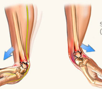
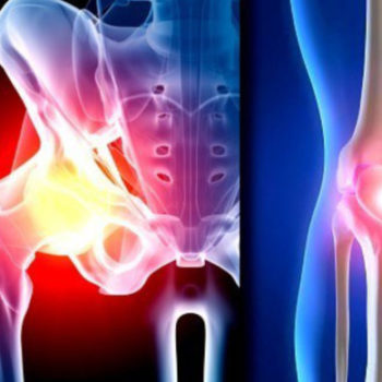
Sarcopenia’s Role in Knee OA Progression

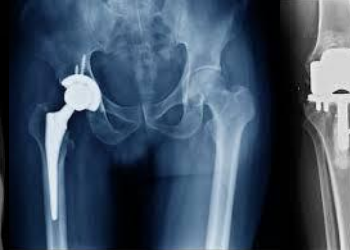
The incidence of total knee arthroplasty to treat end-stage knee osteoarthritis (OA) continues to rise even in the face of patient risk-stratification tools and alternative payment models. Consequently, payers, patients, and their doctors are placing a premium on methods to prolong the native knee joint and delay or avoid surgery. This partly explains the explosion of interest in biologics and the subsequent checkreins being put in place regarding their use.

As the AAOS clinical practice guidelines for the management of knee arthritis clearly state, the best management for symptoms of knee arthritis remains weight loss and self-directed physical activity. However, there is uncertainty regarding which subtypes of patients are likely to achieve OA symptom benefits with different weight-loss strategies.

A recent large, multicenter cohort study published in Arthritis & Rheumatology attempted to further characterize patient body composition and its association with knee OA.  Using whole-body dual x-ray absorptiometry (DXA) measures of fat and muscle mass, researchers classified patients into one of four categories: nonobese nonsarcopenic, sarcopenenic nonobese, nonsarcopenic obese, or sarcopenic obese. Sarcopenia is the general loss of muscle mass associated with aging. If orthopaedic surgeons better understand how fat and muscle metabolism change with time and affect inflammation and chronic disease, they may be able to provide patients with additional insight into preventive measures.

Using DXA-derived calculations, the authors observed that among older adults, the relative risk of developing clinically significant knee osteoarthritis (Kellgren-Lawrence grade ≥2) at 5 years was about 2 times greater in both sarcopenic and nonsarcopenic obese male and female patients compared to nonobese, nonsarcopenic patients.  Sarcopenia alone was not associated with risk of knee OA in women or men. In a sensitivity analysis focusing on BMI, men showed a 3-fold greater risk of knee OA if they were sarcopenic and obese, relative to nonobese nonsarcopenic patients.

The takeaway from this study is that focusing solely on fat/weight loss may overlook a valuable opportunity to slow the progression of knee arthritis in some patients.  Further studies are needed to validate the contribution of low muscle mass to the development and progression of symptomatic knee arthritis.

 Credit to : Jeffrey Stambough, MD(royortho.com)

The role of malnutrition in 90-day outcomes after total joint arthroplasty

Via a cohort study of 4,047 cases, the researchers sought to determine the role of preoperative albumin in total joint arthroplasty (TJA) patients for prediction of length of stay (LOS) and 90-day outcomes. For malnutrition, the albumin cutoff of ≤3.5 g/dL is used as proxy, however the value remained understudied. In some patients at risk for adverse events post TJA, the pre-operative albumin level could be missing. For 90-day readmission, the optimal albumin cutoff was concluded as 3.94 g/dL in a univariable model. Screening for malnutrition as a preoperative evaluation could serve a role. The albumin cutoff value of 3.5 g/dL might be missed.

Read the Full Article on :

https://bit.ly/2QlIuY1

Orthopedic Implants to Dominate the Global 3D Printed Medical Devices Market

Increasing prevalence of chronic diseases and rising awareness regarding personal care will drive future growth.

The global 3D printed medical devices market is expected to expand at a compound annual growth rate (CAGR) of 18.1 percent through 2027. An increasing prevalence of chronic diseases and rising awareness regarding personal care are major factors twill drive growth of the market during this period, according to Future Market Insights.

Increased Ability to Innovate Aids the Global 3D Printed Medical Devices Market
With increasing popularity of 3D printing, medical devices manufacturers are particularly focused on innovations. Substantial need for individualized yet economical medical solutions is met through 3D printing. Complex features of integration such as hard and soft areas, solid and porous structures, multi-material and multi-color, which seemed to be difficult by implementing conventional manufacturing techniques, is made simpler through 3D printing. Patient-specific implants are being manufactured based on computed tomography (CT) and magnetic resonance imaging (MRI) scans provided by surgeons, which results in reduction of the overall surgical cost. This has increased the popularity of 3D printed medical devices all around the world.

The global 3D printed medical devices market is segmented based on application, technology, material type, end user and region. On the basis of application, the market has been segmented into orthopedic Implants, dental implants and cranio-maxillofacial implants. Orthopedic implants application segment accounted for a higher revenue share in global 3D printing devices as compared to others in application segment. Orthopedic implants segment in the application category of the global 3D printed medical devices market was estimated to be valued at nearly $170 million in 2017 and is slated to reach a valuation of nearly $970 million in 2027, exhibiting a 19.2 percent CAGR during the period of assessment.

Based on the material type, 3D printed medical devices market is segmented into metals and alloys, biomaterial inks and plastics. The biomaterial inks segment was estimated to be valued at nearly $65 million in 2017 and is anticipated to reach a valuation of nearly $400 million in 2027, displaying a CAGR of 20 percent during the period of forecast.

Based on the end user, 3D printed medical devices market is segmented into hospitals, ambulatory surgical centres and diagnostic centres. Hospital end user segment accounts for higher demand for 3D printing devices as compared to other distribution channel segments such as ambulatory surgical centres and diagnostic centres, registering a CAGR of 18.7 percent over the forecast period.

On the basis of region, the global 3D printed medical devices market has been segmented into North America, Latin America, Western Europe, Eastern Europe, Asia Pacific excluding Japan, Japan and the Middle East and Africa. Revenues in Western Europe are expected to grow at a CAGR of 18.6 percent, whereas Eastern Europe is anticipated to grow at 15.5 percent over the forecast period. The Western Europe 3D printed medical devices market was estimated to be valued at nearly $65 million in 2017 and is slated to reach a value of nearly $360 million in 2027. The Eastern Europe 3D printed medical devices market was estimated to be valued at nearly $40 million in 2017 and is anticipated to reach a value of nearly $160 millin in 2027.

Some of the players in the global 3D printed medical devices market include 3D Systems Inc., Arcam AB, Stratasys Ltd., FabRx Ltd., EOS GmbH Electro Optical Systems, EnvisionTEC, Cyfuse Biomedical K.K. and Formlabs Inc., among others. Key players are focusing on strengthening their position by establishing new facilities in North America region. Moreover, companies are targeting Asia-Pacific region and Europe by distribution agreements with local players. In order to increase their revenue, major players in the North America region are entering into agreements with hospitals and academic institutes.

Future Market Insights provides market intelligence and consulting services, serving clients in over 150 countries. FMI is headquartered in London and has delivery centers in the United States and India.

From : www.odtmag.com

Efficacy and tolerability of duloxetine in patients with knee osteoarthritis

Researchers assessed the effectiveness and tolerability of duloxetine in patients with knee osteoarthritis (OA), one of the most common joint diseases. Data from six randomized controlled trials were pooled, including 2059 participants. According to findings, duloxetine is effective in managing chronic pain and physical function loss in knee OA with acceptable adverse events despite having no advantage in the treatment of joint rigidity.


Read the Full Article On :

https://www.mdlinx.com/journal-summaries/knee-osteoarthritis-duloxetine-meta-analysis-randomized/2019/05/08/7564361?spec=orthopedics

In MDLinx (A meta-analysis of randomized controlled trials)

What is a Distal Radius Fracture?

The radius is one of two forearm bones and is located on the thumb side. The part of the radius connected to the wrist joint is called the distal radius. When the radius breaks near the wrist, it is called a distal radius fracture.

The break usually happens due to falling on an outstretched or flexed hand. It can also happen in a car accident, a bike accident, a skiing accident or another sports activity.

A distal radius fracture can be isolated, which means no other fractures are involved. It can also occur along with a fracture of the distal ulna (the forearm bone on the small finger side). In these cases, the injury is called a distal radius and ulna fracture.

Depending on the angle of the distal radius as it breaks, the fracture is called a Colles or Smith fracture.

  • Colles fracture may result from direct impact to the palm, like if you use your hands to break up a fall and land on the palms. The side view of a wrist after a Colles fracture is sometimes compared to the shape of a fork facing down. There is a distinct “bump” in the wrist similar to the neck of the fork. It happens because the broken end of the distal radius shifts up toward the back of the hand.
  • Smith fracture is the less common of the two. It may result from an impact to the back of the wrist, such as falling on a bent wrist. The end of the distal radius typically shifts down toward the palm side in this type of fracture. This usually makes for a distinct drop in the wrist where the longer part of the radius ends.

What are the symptoms of a distal radius fracture?

  • Immediate pain with tenderness when touched
  • Bruising and swelling around the wrist
  • Deformity — the wrist being in an odd position

What is the treatment for a distal radius fracture?

Decisions on how to treat a distal radius fracture may depend on many factors, including:

  • Fracture displacement (whether the broken bones shifted)
  • Comminution (whether there are fractures in multiple places)
  • Joint involvement
  • Associated ulna fracture and injury to the median nerve
  • Whether it is the dominant hand
  • Your occupation and activity level

Surgery for Distal Radius Fractures

This option is usually for fractures that are considered unstable or can’t be treated with a cast. Surgery is typically performed through an incision over the volar aspect of your wrist (where you feel your pulse). This allows full access to the break. The pieces are put together and held in place with one or more plates and screws.

In certain cases, a second incision is required on the back side of your wrist to re-establish the anatomy. Plates and screws will be used to hold the pieces in place. If there are multiple bone pieces, fixation with plates and screws may not be possible. In these cases, an external fixator with or without additional wires may be used to secure the fracture. With an external fixator, most of the hardware remains outside of the body.

After the surgery, a splint will be placed for two weeks until your first follow-up visit. At that time, the splint will be removed and exchanged with a removable wrist splint. You will have to wear it for four weeks. You will start your physical therapy to regain wrist function and strength after your first clinic visit. Six weeks after your surgery, you may stop wearing the removable splint. You should continue the exercises prescribed by your surgeon and therapist. Early motion is key to achieving the best recovery after surgery.

Written By : Johns Hopkins Medicine

Clavicle Fracture

A clavicle fracture is a break in the collarbone, one of the main bones in the shoulder. This type of fracture is fairly common—accounting for about 5 percent of all adult fractures. Most clavicle fractures occur when a fall onto the shoulder or an outstretched arm puts enough pressure on the bone that it snaps or breaks. A broken collarbone can be very painful and can make it hard to move your arm.Most clavicle fractures can be treated by wearing a sling to keep the arm and shoulder from moving while the bone heals. With some clavicle fractures, however, the pieces of bone move far out of place when the injury occurs. For these more complicated fractures, surgery may be needed to realign the collarbone.

Anatomy

The clavicle is located between the ribcage (sternum) and the shoulder blade (scapula). It is the bone that connects the arm to the body.

The clavicle lies above several important nerves and blood vessels. However, these vital structures are rarely injured when a fracture occurs.

Description

Clavicle fractures are fairly common and occur in people of all ages. Most fractures occur in the middle portion, or shaft, of the bone. Occasionally, the bone will break where it attaches at the ribcage or shoulder blade.

Clavicle fractures vary. The bone can crack just slightly or break into many pieces (comminuted fracture). The broken pieces of bone may line up straight or may be far out of place (displaced fracture).

Cause

Clavicle fractures are most often caused by a direct blow to the shoulder. This can happen during a fall onto the shoulder or a car collision. A fall onto an outstretched arm can also cause a clavicle fracture. In a baby, a clavicle fracture can occur during the passage through the birth canal.

Symptoms

A clavicle fracture can be very painful and may make it hard to move your arm. Other signs and symptoms of a fracture may include:

  • Sagging of the shoulder downward and forward
  • Inability to lift the arm because of pain
  • A grinding sensation when you try to raise the arm
  • A deformity or “bump” over the break
  • Bruising, swelling, and/or tenderness over the collarbone

Femur Shaft Fractures (Broken Thighbone)

Our thighbone (femur) is the longest and strongest bone in our body. Because the femur is so strong, it usually takes a lot of force to break it. Motor vehicle collisions, for example, are the number one cause of femur fractures.

The long, straight part of the femur is called the femoral shaft. When there is a break anywhere along this length of bone, it is called a femoral shaft fracture. This type of broken leg almost always requires surgery to heal.

Types of Femoral Shaft Fractures

Femur fractures vary greatly, depending on the force that causes the break. The pieces of bone may line up correctly (stable fracture) or be out of alignment (displaced fracture). The skin around the fracture may be intact (closed fracture) or the bone may puncture the skin (open fracture).

Doctors describe fractures to each other using classification systems. Femur fractures are classified depending on:

  • The location of the fracture (the femoral shaft is divided into thirds: distal, middle, proximal)
  • The pattern of the fracture (for example, the bone can break in different directions, such as crosswise, lengthwise, or in the middle)
  • Whether the skin and muscle over the bone is torn by the injury

The most common types of femoral shaft fractures include:

Transverse fracture. In this type of fracture, the break is a straight horizontal line going across the femoral shaft.

Oblique fracture. This type of fracture has an angled line across the shaft.

Spiral fracture. The fracture line encircles the shaft like the stripes on a candy cane. A twisting force to the thigh causes this type of fracture.

Comminuted fracture. In this type of fracture, the bone has broken into three or more pieces. In most cases, the number of bone fragments corresponds with the amount of force needed to break the bone.

Open fracture. If a bone breaks in such a way that bone fragments stick out through the skin or a wound penetrates down to the broken bone, the fracture is called an open or compound fracture. Open fractures often involve much more damage to the surrounding muscles, tendons, and ligaments. They have a higher risk for complications—especially infections—and take a longer time to heal.

Cause

Femoral shaft fractures in young people are frequently due to some type of high-energy collision. The most common cause of femoral shaft fracture is a motor vehicle or motorcycle crash. Being hit by a car while walking is another common cause, as are falls from heights and gunshot wounds.

A lower-force incident, such as a fall from standing, may cause a femoral shaft fracture in an older person who has weaker bones.

Symptoms

A femoral shaft fracture usually causes immediate, severe pain. You will not be able to put weight on the injured leg, and it may look deformed—shorter than the other leg and no longer straight.

Credit to: orthoinfo

New Data Links High Volume TJA To Patient Outcomes

Researchers at the University of Rochester in New York have taken a deep look into the connection between the volume of revision total joint arthroplasty (TJA) surgeries and patient outcomes.

Co-author Benjamin Ricciardi, M.D. explained to OTW why this connection is especially important. “The association between hospital volume and primary total joint replacement has been well established but has not been studied extensively in revision total joint replacement. Revision surgery is more complicated than primary surgery, and we hypothesized that hospitals with higher volume would have improved early outcomes relative to lower volume hospitals.”

The researchers looked at records from 29,948 inpatient stays for revision TJA from 2008 to 2014 in the Statewide Planning and Research Cooperative System (SPARCS) database for New York State. They looked at the relationship between hospital revision volumes by quartile and the associated patient outcomes. The researchers also examined the top 5 percentile of hospitals as a separate cohort.

Dr. Ricciardi summarized the study results to OTW, “The most important results were hospitals in the highest 5% by volume for revision total joint replacement had lower 90-day readmissions relative to lower volume hospitals. Additionally, hospitals in the lowest quartile by volume had higher 90-day complications relative to higher volume hospital categories.”

“The results of this study suggest that regionalizing revision services to higher volume hospitals may be beneficial to early revision total joint replacement outcomes, however, further studies are needed to examine any negative impact on access to care that these policies may cause and define longer term outcomes of revision total joint replacement relative to hospital volume.”

According to the authors, “Disadvantages of regionalization include reduced access to care, increased patient travel distances, and possible capacity issues at receiving centers. Further studies are needed to evaluate the benefits and negative consequences of regionalizing revision TJA services to higher-volume revision TJA institutions.”


Reference from : ryortho.com

New Protocol May Decrease Hip Dislocation Risk

Researchers from NYU Langone Orthopedics, Hospital for Special Surgery, and Mayo Clinic have developed a hip-spine classification system for use in revision total hip arthroplasty that may help decrease the risk of recurrent instability.

Their work, “Evaluation of the Spine is Critical in Patients with Recurrent Instability after Total Hip Arthroplasty,” has been accepted for publication in an upcoming 2019 issue of The Bone & Joint Journal.

Co-author Jonathan M. Vigdorchik, M.D., at the time of the study an orthopedic surgeon in the Division of Adult Reconstructive Surgery at NYU Langone Orthopedics in New York City, noticed a trend which, as he explained to OTW, prompted this study. “In clinical practice, being at a tertiary referral center, we get many referrals for revision THA [total hip arthroplasty]. As we started looking at our revisions for dislocation, we began noticing a trend—patients all had spine fusions or spines that were in bad shape. So, this launched us on a research path about spine fusions and hip replacement.”

The researchers collected data on 111 patients undergoing revision THA for recurrent instability and matched 1:1 to 111 revisions specifically performed for instability not using this protocol (the control group).

Dr. Vigdorchik, now an orthopaedic hip and knee replacement surgeon at Hospital for Special Surgery in New York, told OTW, “We found that spinal fusion, and also the higher number of spinal levels fused, caused higher dislocation rates. We also found that patients with spinal deformity had higher rates of dislocation.”

“So, we had a combined spine surgeons and hip surgeons conference to discuss. As a hip surgeon, I started looking at the hip and the pelvis like a spine surgeon and began noticing certain trends. When we applied this to dislocating hips, it became very clear why they were dislocating and explains a large group of patients where the doctors could never figure out why the X-rays looked good (or so they thought).

“But they were just looking at the wrong X-rays. So that is why I came up with this protocol, to teach them the right X-rays to do and then what to do with what they found.”

“Revising a hip that is dislocating has a really high complication rate, especially recurrent dislocation.

From : www.ryortho.com/

Outcomes of superficial and deep irrigation and debridement in total hip and knee arthroplasty

Researchers performed a retrospective study of 176 patients. that underwent irrigation and debridement(I&D) within 28 days of  TJA, to examine the role and outcomes of both superficial and deep I&D in patients with wound-related issues and/or suspected periprosthetic joint infection (PJI). For superficial I&D, the overall success was 84.28% compared to 68.86% for deep I&D. Outcomes support the viability of superficial I&D in patients with wound-related issues as long as joint aspiration is performed to rule out infection involving the prosthesis. They suggest opening fascia and exploring deeper tissues in case there are findings of no fluid or purulence.


Read the Full Article On : https://www.arthroplastyjournal.org/article/S0883-5403(19)30255-4/fulltext?rss=yes

    We offer a wide variety of safe and advanced Orthopedic Implants and fixators. At the helm of the company’s operations is Mr. Vinodbhai, the Director. His enterprising skills and experience in the industry continue to play a pivotal role in the company’s growth.We are based in Ahmedabad city West Part of India.

    ADDRESS

    Plot No.10, Phase-1, B|h. Prashant Eng., G.I.D.C.Vatva, Ahmedabad-382 445, Gujarat, (INDIA).

    PHONE

    +91 9375801932

    EMAIL

    [email protected]

    Cart