Tag Archives: patient

Efficacy and tolerability of duloxetine in patients with knee osteoarthritis

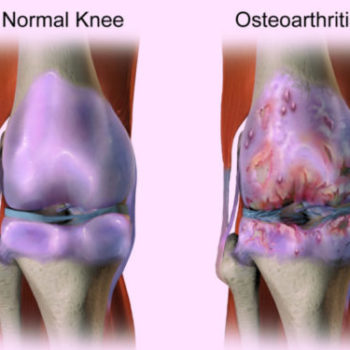
Researchers assessed the effectiveness and tolerability of duloxetine in patients with knee osteoarthritis (OA), one of the most common joint diseases. Data from six randomized controlled trials were pooled, including 2059 participants. According to findings, duloxetine is effective in managing chronic pain and physical function loss in knee OA with acceptable adverse events despite having no advantage in the treatment of joint rigidity.


Read the Full Article On :

https://www.mdlinx.com/journal-summaries/knee-osteoarthritis-duloxetine-meta-analysis-randomized/2019/05/08/7564361?spec=orthopedics

In MDLinx (A meta-analysis of randomized controlled trials)

What is a Distal Radius Fracture?

The radius is one of two forearm bones and is located on the thumb side. The part of the radius connected to the wrist joint is called the distal radius. When the radius breaks near the wrist, it is called a distal radius fracture.

The break usually happens due to falling on an outstretched or flexed hand. It can also happen in a car accident, a bike accident, a skiing accident or another sports activity.

A distal radius fracture can be isolated, which means no other fractures are involved. It can also occur along with a fracture of the distal ulna (the forearm bone on the small finger side). In these cases, the injury is called a distal radius and ulna fracture.

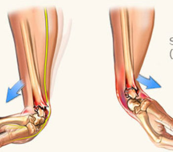
Depending on the angle of the distal radius as it breaks, the fracture is called a Colles or Smith fracture.

  • Colles fracture may result from direct impact to the palm, like if you use your hands to break up a fall and land on the palms. The side view of a wrist after a Colles fracture is sometimes compared to the shape of a fork facing down. There is a distinct “bump” in the wrist similar to the neck of the fork. It happens because the broken end of the distal radius shifts up toward the back of the hand.
  • Smith fracture is the less common of the two. It may result from an impact to the back of the wrist, such as falling on a bent wrist. The end of the distal radius typically shifts down toward the palm side in this type of fracture. This usually makes for a distinct drop in the wrist where the longer part of the radius ends.

What are the symptoms of a distal radius fracture?

  • Immediate pain with tenderness when touched
  • Bruising and swelling around the wrist
  • Deformity — the wrist being in an odd position

What is the treatment for a distal radius fracture?

Decisions on how to treat a distal radius fracture may depend on many factors, including:

  • Fracture displacement (whether the broken bones shifted)
  • Comminution (whether there are fractures in multiple places)
  • Joint involvement
  • Associated ulna fracture and injury to the median nerve
  • Whether it is the dominant hand
  • Your occupation and activity level

Surgery for Distal Radius Fractures

This option is usually for fractures that are considered unstable or can’t be treated with a cast. Surgery is typically performed through an incision over the volar aspect of your wrist (where you feel your pulse). This allows full access to the break. The pieces are put together and held in place with one or more plates and screws.

In certain cases, a second incision is required on the back side of your wrist to re-establish the anatomy. Plates and screws will be used to hold the pieces in place. If there are multiple bone pieces, fixation with plates and screws may not be possible. In these cases, an external fixator with or without additional wires may be used to secure the fracture. With an external fixator, most of the hardware remains outside of the body.

After the surgery, a splint will be placed for two weeks until your first follow-up visit. At that time, the splint will be removed and exchanged with a removable wrist splint. You will have to wear it for four weeks. You will start your physical therapy to regain wrist function and strength after your first clinic visit. Six weeks after your surgery, you may stop wearing the removable splint. You should continue the exercises prescribed by your surgeon and therapist. Early motion is key to achieving the best recovery after surgery.

Written By : Johns Hopkins Medicine

Clavicle Fracture

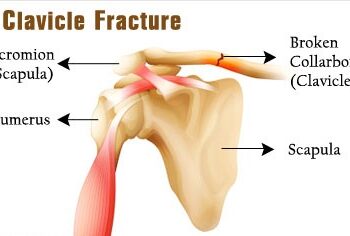
A clavicle fracture is a break in the collarbone, one of the main bones in the shoulder. This type of fracture is fairly common—accounting for about 5 percent of all adult fractures. Most clavicle fractures occur when a fall onto the shoulder or an outstretched arm puts enough pressure on the bone that it snaps or breaks. A broken collarbone can be very painful and can make it hard to move your arm.Most clavicle fractures can be treated by wearing a sling to keep the arm and shoulder from moving while the bone heals. With some clavicle fractures, however, the pieces of bone move far out of place when the injury occurs. For these more complicated fractures, surgery may be needed to realign the collarbone.

Anatomy

The clavicle is located between the ribcage (sternum) and the shoulder blade (scapula). It is the bone that connects the arm to the body.

The clavicle lies above several important nerves and blood vessels. However, these vital structures are rarely injured when a fracture occurs.

Description

Clavicle fractures are fairly common and occur in people of all ages. Most fractures occur in the middle portion, or shaft, of the bone. Occasionally, the bone will break where it attaches at the ribcage or shoulder blade.

Clavicle fractures vary. The bone can crack just slightly or break into many pieces (comminuted fracture). The broken pieces of bone may line up straight or may be far out of place (displaced fracture).

Cause

Clavicle fractures are most often caused by a direct blow to the shoulder. This can happen during a fall onto the shoulder or a car collision. A fall onto an outstretched arm can also cause a clavicle fracture. In a baby, a clavicle fracture can occur during the passage through the birth canal.

Symptoms

A clavicle fracture can be very painful and may make it hard to move your arm. Other signs and symptoms of a fracture may include:

  • Sagging of the shoulder downward and forward
  • Inability to lift the arm because of pain
  • A grinding sensation when you try to raise the arm
  • A deformity or “bump” over the break
  • Bruising, swelling, and/or tenderness over the collarbone

New Data Links High Volume TJA To Patient Outcomes

Researchers at the University of Rochester in New York have taken a deep look into the connection between the volume of revision total joint arthroplasty (TJA) surgeries and patient outcomes.

Co-author Benjamin Ricciardi, M.D. explained to OTW why this connection is especially important. “The association between hospital volume and primary total joint replacement has been well established but has not been studied extensively in revision total joint replacement. Revision surgery is more complicated than primary surgery, and we hypothesized that hospitals with higher volume would have improved early outcomes relative to lower volume hospitals.”

The researchers looked at records from 29,948 inpatient stays for revision TJA from 2008 to 2014 in the Statewide Planning and Research Cooperative System (SPARCS) database for New York State. They looked at the relationship between hospital revision volumes by quartile and the associated patient outcomes. The researchers also examined the top 5 percentile of hospitals as a separate cohort.

Dr. Ricciardi summarized the study results to OTW, “The most important results were hospitals in the highest 5% by volume for revision total joint replacement had lower 90-day readmissions relative to lower volume hospitals. Additionally, hospitals in the lowest quartile by volume had higher 90-day complications relative to higher volume hospital categories.”

“The results of this study suggest that regionalizing revision services to higher volume hospitals may be beneficial to early revision total joint replacement outcomes, however, further studies are needed to examine any negative impact on access to care that these policies may cause and define longer term outcomes of revision total joint replacement relative to hospital volume.”

According to the authors, “Disadvantages of regionalization include reduced access to care, increased patient travel distances, and possible capacity issues at receiving centers. Further studies are needed to evaluate the benefits and negative consequences of regionalizing revision TJA services to higher-volume revision TJA institutions.”


Reference from : ryortho.com

New Protocol May Decrease Hip Dislocation Risk

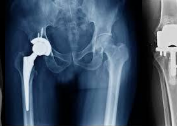
Researchers from NYU Langone Orthopedics, Hospital for Special Surgery, and Mayo Clinic have developed a hip-spine classification system for use in revision total hip arthroplasty that may help decrease the risk of recurrent instability.

Their work, “Evaluation of the Spine is Critical in Patients with Recurrent Instability after Total Hip Arthroplasty,” has been accepted for publication in an upcoming 2019 issue of The Bone & Joint Journal.

Co-author Jonathan M. Vigdorchik, M.D., at the time of the study an orthopedic surgeon in the Division of Adult Reconstructive Surgery at NYU Langone Orthopedics in New York City, noticed a trend which, as he explained to OTW, prompted this study. “In clinical practice, being at a tertiary referral center, we get many referrals for revision THA [total hip arthroplasty]. As we started looking at our revisions for dislocation, we began noticing a trend—patients all had spine fusions or spines that were in bad shape. So, this launched us on a research path about spine fusions and hip replacement.”

The researchers collected data on 111 patients undergoing revision THA for recurrent instability and matched 1:1 to 111 revisions specifically performed for instability not using this protocol (the control group).

Dr. Vigdorchik, now an orthopaedic hip and knee replacement surgeon at Hospital for Special Surgery in New York, told OTW, “We found that spinal fusion, and also the higher number of spinal levels fused, caused higher dislocation rates. We also found that patients with spinal deformity had higher rates of dislocation.”

“So, we had a combined spine surgeons and hip surgeons conference to discuss. As a hip surgeon, I started looking at the hip and the pelvis like a spine surgeon and began noticing certain trends. When we applied this to dislocating hips, it became very clear why they were dislocating and explains a large group of patients where the doctors could never figure out why the X-rays looked good (or so they thought).

“But they were just looking at the wrong X-rays. So that is why I came up with this protocol, to teach them the right X-rays to do and then what to do with what they found.”

“Revising a hip that is dislocating has a really high complication rate, especially recurrent dislocation.

From : www.ryortho.com/

Outcomes of superficial and deep irrigation and debridement in total hip and knee arthroplasty

Researchers performed a retrospective study of 176 patients. that underwent irrigation and debridement(I&D) within 28 days of  TJA, to examine the role and outcomes of both superficial and deep I&D in patients with wound-related issues and/or suspected periprosthetic joint infection (PJI). For superficial I&D, the overall success was 84.28% compared to 68.86% for deep I&D. Outcomes support the viability of superficial I&D in patients with wound-related issues as long as joint aspiration is performed to rule out infection involving the prosthesis. They suggest opening fascia and exploring deeper tissues in case there are findings of no fluid or purulence.


Read the Full Article On : https://www.arthroplastyjournal.org/article/S0883-5403(19)30255-4/fulltext?rss=yes

Time to seriously consider focused mris for injured spines

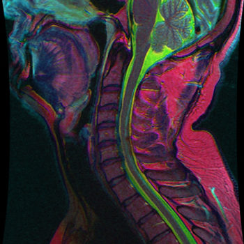
Is it sufficient to only examine the injured part of the spine via focused magnetic resonance imaging (FMRI) or will that lead to missed problems?

In patients who sustain blunt trauma, CT is the initial screening modality of choice in most emergency departments in the United States to look for injury to various parts of the body—including the spine. If a fracture is seen in the thoracic or lumbar spine on CT, sometimes an MRI of the injured segment of the spine is useful to provide additional information about the injury to help physicians decide on a treatment plan.”

“The problem is that MRI is a relatively expensive and time-consuming test compared to CT, so every time we were ordering MRIs of the entire spine in this scenario, it seemed somewhat excessive. Since we are mostly interested in looking at the part of the spine that we already know is injured (based on the CT), we asked ourselves if just imaging the injured portion of the spine by MRI (a focused MRI) would be adequate to provide the information we need to decide on a treatment plan. The concern with this approach is whether we would miss injuries by not looking at the entire spine by MRI.”

To answer the question, the investigators reviewed records for all adult trauma patients who presented to Mass General’s emergency department between 2008 and 2016 with one or more fractures of the thoracic and/or lumbar spine—as identified using computed tomography (CT) and who underwent MRI of the entire thoracic and lumbar spine within 10 days.

The most important result of the test, said Dr. Karim, was “that performing a focused MRI of only the injured portion of the spine would lead physicians to missing some sort of injury in another portion of the spine about 15% of the time, but none of these ‘missed injuries’ would lead to any change in treatment plan or patient care if they are known.”

“This is a very practical study from our perspective because it affects the day-to-day work of radiologists, emergency room physicians, and spine surgeons. If they see a fracture in the thoracic or lumbar spine on CT that they want to better characterize on MRI, they should be comfortable obtaining just a focused MRI of the injured portion of the spine—saving time, money, and patient discomfort.”

Reference From : https://ryortho.com

Patient relevant outcomes of unicompartmental vs total knee replacement

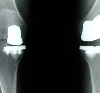
Between 1 January 1997 and 31 December 2018, via searching Medline, Embase, Cochrane Controlled Register of Trials (CENTRAL), and Clinical Trials.gov, researchers provided a clear and comprehensive summary of the published data on unicompartmental knee replacement (UKA) or total knee replacement (TKA), comparing domains of outcome that have been shown to be important to patients and clinicians in order to enable informed decision making. Findings suggested that TKA and UKA are both viable treatment options for isolated unicompartmental osteoarthritis arthritis. This investigation shows better results for UKA in several outcome domains by directly comparing the two treatments. The risk of revision surgery for TKA, however, was lower. This information should be available to patients when selecting treatment options as part of the shared decision-making process.

Read the Full Article : https://www.bmj.com/content/364/bmj.l352

Reference From : www.mdlinx.com

    We offer a wide variety of safe and advanced Orthopedic Implants and fixators. At the helm of the company’s operations is Mr. Vinodbhai, the Director. His enterprising skills and experience in the industry continue to play a pivotal role in the company’s growth.We are based in Ahmedabad city West Part of India.

    ADDRESS

    Plot No.10, Phase-1, B|h. Prashant Eng., G.I.D.C.Vatva, Ahmedabad-382 445, Gujarat, (INDIA).

    PHONE

    +91 9375801932

    EMAIL

    [email protected]

    Cart