Tag Archives: orthopaedic implants company

You know what is the Distraction osteogenesis?

Distraction osteogenesis (DO), also called callus distractioncallotasis and osteodistraction, is a process used in orthopedic surgery, podiatric surgery, and oral and maxillofacial surgery to repair skeletal deformities and in reconstructive surgery. The procedure involves cutting and slowly separating bone, allowing the bone healing process to fill in the gap.

Medical Uses

Distraction osteogenesis (DO) is used in orthopedic surgery, podiatric surgery, and oral and maxillofacial surgery to repair skeletal deformities and in reconstructive surgery. It was originally used to treat problems like unequal leg length, but since the 1980s is most commonly used to treat issues like hemifacial microsomia, micrognathism (chin so small it causes health problems), craniofrontonasal dysplasias, craniosynostosis, as well as airway obstruction in babies caused by glossoptosis (tongue recessed too far back in the mouth) or micrognathism.

In 2016 a systematic review of papers describing bone and soft tissue outcomes of DO procedures on the lower jawbone was published; the authors had planned to do a meta-analysis but found the studies were too poor in quality and too heterogeneous to pool. From what they were able to generalize, the authors found there was significant relapse in the vertical plane for bone, and a higher risk of relapse when there was an initial high gonial angle or Jarabak ratio (sella–gonion/nasion–menton). For soft tissue, little evidence was available regarding the vertical dimension, while a 90% correspondence between skeletal and soft tissue was found for sagittal positioning; the dental-to-soft tissue agreement was around 20%.

A 2016 Cochrane review of DO on the upper jawbone to treat cleft lip and cleft palate compared with orthognathic surgery found only one study, involving 47 participants and performed between 2002 and 2008 at the University of Hong Kong. This was not sufficient evidence from which to generalize, but the authors noted that while both procedures produced notable hard and soft tissue improvements, the DO group had greater advancement of the maxillary and less horizontal relapse five years after surgery. There was no difference in speech or nasal emissions outcomes nor in adverse effects; the DO group had lower satisfaction at three months after surgery but higher at two years.
Distraction osteogenesis

Procedure

In the first phase, called the “osteotomy/surgical phase”, the bone is cut, either partially, only through the hard exterior, or completely, and a device is fitted which will be used in the next phases. In the second phase, the latency period, which lasts generally seven days, the appliance is not activated and early stages of bone healing are allowed. In the third phase, the “distraction phase”, the device, which is mounted to the bone on each side of the cut, is used to gradually separate the two pieces, allowing new bone to form in the gap. When the desired or possible length is reached, which usually takes three to seven days, a consolidation phase follows in which the device keeps the bone stable to allow the bone to fully heal. After the consolidation phase, the device is removed in a second surgical procedure.

The device that is used is usually manually operated by twisting a rod that through a rack and pinion system or the like, separates the bone; the rate of separation is carefully determined because going too quickly can cause nonunion, in which unstable fibrous connective tissue is formed instead of bone, and going too slowly can allow premature union to occur. Generally the rate is about a millimeter per day, achieved in two steps per day. The frequency of steps and how much the device is moved at each step, is called the “rhythm”. The devices sometimes contain a spring that provides tension to continually separate the bones, instead of being manually operated at set intervals.
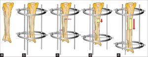
 

Distraction osteogenesis


Despite these manually operated systems there are also motorized systems like the FITBONE from WITTENSTEIN. The FITBONE is a fully implantable, motorized, lengthening and correction nail. Advantages of this device are accurate deformity correction, low scar tissue formation, and reduced risk of infection. Furthermore the patients describe the procedure as more comfortable than limb lengthening with mechanical systems.

Content credit to wikipedia.org

Image credit to researchget.net

Anterior cervical discectomy and fusion(ACDF)

Anterior cervical discectomy and fusion (ACDF) is a surgical procedure to treat nerve root or spinal cord compression by decompressing the spinal cord and nerve roots of the cervical spine with a discectomy, followed by inter-vertebral fusion to stabilize the corresponding vertebrae.This procedure is used when other non-surgical treatments have failed.

Medical uses

ACDF is used to treat serious pain from a nerve root  that has become inflamed. This can be caused by:

1. a herniated disc when other non-surgical treatments have failed. The nucleus pulposus (the jelly-like center of the disc) of the herniated disc bulges out through the annulus (surrounding wall) and presses on the nerve root next to it.

2. degenerative disc disease (spondylosis). The disc consists of about 80% water. When one grows older, the disc starts to dry out and shrink, causing small tears in the annulus and inflammation of the nerve root.


Technique

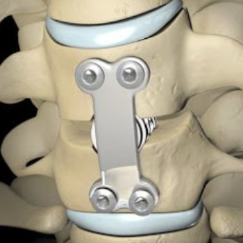
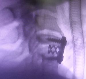
Anterior cervical discectomy and fusion(ACDF) surgery

The neurosurgeon or orthopedic surgeon enters the space between two discs through a small incision in front (= anterior) of and at the right or left side of the neck. The disc is completely removed, as well as arthritic bone spurs. The disc material, pressing on the spinal nerve or spinal cord, is then completely removed. The intervertebral foramen, the bone channel through which the spinal nerve runs, is then enlarged with a drill giving the nerve more room to exit the spinal canal.

To prevent the vertebrae from collapsing and to increase stability, the open space is often filled with a graft. That can be a bone graft, taken from the pelvis or cadaveric bone; or an artificial implant. The slow process of the bone graft joining the vertebrae together is called “fusion”. Sometimes a titanium plate is screwed on the vertebrae or screws are used between the vertebrae to increase stability during fusion, especially when there is more than one disc involved.

Anterior cervical discectomy and fusion(ACDF)  surgery


Recovery

The surgery requires a short stay in the clinic (1 to 3 days) and a gradual recovery between 1 and 6 weeks. However, the technology has advanced and it can be performed by ‘Endoscopic Micro Discectomy” with the patient able to continue their normal life in two days. The patient may be advised to wear a neck brace or collar (for up to 8 weeks) that serves to ensure proper spinal alignment. Wearing the brace heightens one’s awareness of posture and positioning and helps prevent movements (e.g., sudden and/or excessive bending or twisting of the neck) that may aggravate or slow down the healing process.

It is especially advisable to wear a protective neck brace when traveling (e.g., by car), sleeping, showering, or any other activities in which the patient may not be able to be ensure proper spinal alignment. In addition, physical therapy and related healing modalities (e.g., massage, acupuncture) may be recommended in order to promote proper healing, as well as to strengthen the surrounding muscles that can take over the neck brace’s ‘job’ of ensuring proper spinal alignment when the patient starts (around 4 to 6 weeks after surgery) to wean off the neck brace.

Article From : wikipedia.com

Sarcopenia’s Role in Knee OA Progression

The incidence of total knee arthroplasty to treat end-stage knee osteoarthritis (OA) continues to rise even in the face of patient risk-stratification tools and alternative payment models. Consequently, payers, patients, and their doctors are placing a premium on methods to prolong the native knee joint and delay or avoid surgery. This partly explains the explosion of interest in biologics and the subsequent checkreins being put in place regarding their use.

As the AAOS clinical practice guidelines for the management of knee arthritis clearly state, the best management for symptoms of knee arthritis remains weight loss and self-directed physical activity. However, there is uncertainty regarding which subtypes of patients are likely to achieve OA symptom benefits with different weight-loss strategies.

A recent large, multicenter cohort study published in Arthritis & Rheumatology attempted to further characterize patient body composition and its association with knee OA.  Using whole-body dual x-ray absorptiometry (DXA) measures of fat and muscle mass, researchers classified patients into one of four categories: nonobese nonsarcopenic, sarcopenenic nonobese, nonsarcopenic obese, or sarcopenic obese. Sarcopenia is the general loss of muscle mass associated with aging. If orthopaedic surgeons better understand how fat and muscle metabolism change with time and affect inflammation and chronic disease, they may be able to provide patients with additional insight into preventive measures.

Using DXA-derived calculations, the authors observed that among older adults, the relative risk of developing clinically significant knee osteoarthritis (Kellgren-Lawrence grade ≥2) at 5 years was about 2 times greater in both sarcopenic and nonsarcopenic obese male and female patients compared to nonobese, nonsarcopenic patients.  Sarcopenia alone was not associated with risk of knee OA in women or men. In a sensitivity analysis focusing on BMI, men showed a 3-fold greater risk of knee OA if they were sarcopenic and obese, relative to nonobese nonsarcopenic patients.

The takeaway from this study is that focusing solely on fat/weight loss may overlook a valuable opportunity to slow the progression of knee arthritis in some patients.  Further studies are needed to validate the contribution of low muscle mass to the development and progression of symptomatic knee arthritis.

 Credit to : Jeffrey Stambough, MD(royortho.com)

Type of Knee Ligament Injuries

Our knee is made up of many important structures, any of which can be injured. The most common knee injuries include fractures around the knee, dislocation, and sprains and tears of soft tissues, like ligaments. In many cases, injuries involve more than one structure in the knee.

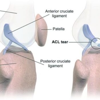
Anterior Cruciate Ligament (ACL) Injuries

The anterior cruciate ligament is often injured during sports activities. Athletes who participate in high demand sports like soccer, football, and basketball are more likely to injure their anterior cruciate ligaments. Changing direction rapidly or landing from a jump incorrectly can tear the ACL. About half of all injuries to the anterior cruciate ligament occur along with damage to other structures in the knee, such as articular cartilage, meniscus, or other ligaments.

Posterior Cruciate Ligament (PCL) Injuries

The posterior cruciate ligament is often injured from a blow to the front of the knee while the knee is bent. This often occurs in motor vehicle crashes and sports-related contact. Posterior cruciate ligament tears tend to be partial tears with the potential to heal on their own.

ROSACON 2019

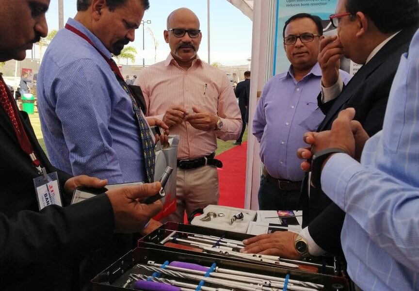
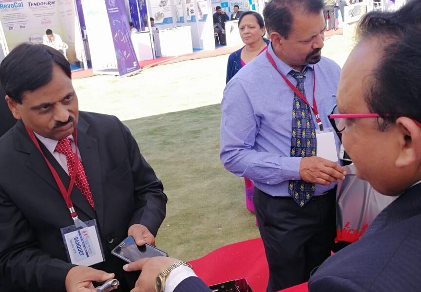
 31st ROSACON-2019 to be held at Labh Garh Resort, Udaipur From 15th to 17th Feb 2019.

We exhibited all our Orthopaedic Implants(Products) & Instruments.


UPORTHOCON 2019

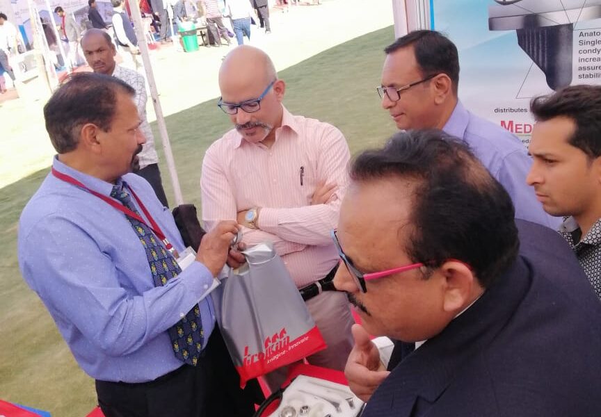
We participated in UPORTHOCON  2019 43th Annual Conference of UP Orthopaedic Association held in UP from 15th to 17th  February, 2019 at Rohilkhand Medical college, Bareilly.

We exhibited all our Articular and Periarticular Trauma Implants (products), Total Joint Replacement System & Instruments.

Exercise After Hip Replacement

To achieve full recovery after a hip replacement it is vital that you incorporate regular exercise into your life. Regular post-operative exercise will allow you to return to your everyday activities within 3-6 weeks after surgery; and return to driving at six weeks. These exercises are geared to restoring your blood flow, strength and mobility. Moving forward it is important to gradually increase walking, sitting, standing, and climbing stairs.

Your orthopedic surgeon will work with your physical therapist to create a plan for you.

During early recovery, while you are in the hospital, you will begin to walk short distances in your room to help your hip to regain its strength and movement.

Post- operative exercise

Walking

Walking is the best exercise for a healthy recovery, because walking will help you recover hip movement. Initially, the use of a walker or crutches will help to prevent blood clots and strengthen your muscles which will improve hip movement. To ensure you are walking properly you will receive guidance from your surgeon or therapist about how much weight to put on the leg. You will be able to more weight on your operated leg as your strength and endurance improve.

While exercise may be painful at first, it can reduce post-op pain, improve blood flow and speed recovery. This will also reduce swelling in the calf and ankle. Swelling can last up to 3 months.

We recommend that you walk two to three times a day for about 20-30 minutes each time. You should get up and walk around the house every 1-2 hours.  Eventually you will be able to walk and stand for more than 10 minutes without putting weight on your walker or crutches. Then you can graduate to a cane.

Climbing Stairs

Stair climbing is a great way to increase your strength and endurance. Always use your hand rail and do not try to climb any steps that are higher than 7″. Using a crutch on the opposite side from your surgery, climb up leading with your good leg. Putting weight on the crutch, raise your operated leg and place it on the step. Moving slowly one step at a time.

Going down lead with your operated leg, putting your weight on the crutch. Eventually, you will become stronger enabling you to climb the stairs foot over foot.

Early Post-op exercises

Lying Down:

  1. Ankle Pumps- This exercise should be done right after surgery, and until you are completely recovered. Point and flex your ankles often, at least once per hour.
  2. Ankle Rotations- move the ankle inside and outside away from the other foot. Do these 5 times in each direction, 3 to 4 times per day.
  3. Knee Bends – lying on the bed with your leg straight out in front of you, pull your foot toward your buttocks keeping your heel on the bed. Hold in this position for 5-10 seconds, then straighten the knee keeping your heel on the bed.
  4. Buttock Tightening- Lying on your back, contract your buttocks muscles and hold for a count of 5. Release and repeat 10 times a day.
  5. Abduction exercises- slide your leg out to your side as far as you can away from your body, and then slide it back. Repeat 10 times per day.
  6. Quadriceps – Lying on your back, tighten your thigh muscles. Try to straighten your knee. Hold for 5-10 seconds. Repeat 10 reps in 10 minutes, resting one minute, then repeat. Stop when your thigh feels fatigued.
  7. Straight leg raises- tighten your quads keeping your knee straight. Lift the leg a few inches. hold 5-10 seconds. Lower the leg. Repeat until your thigh feels fatigued.

Standing Exercises

  1. Knee raises. Standing behind a chair and hold the back of the chair for support, raise the knee toward your chest only to waist height. Hold for a count of 2-3 and put the leg down.
  2. Standing hip abduction. Holding the back of a chair for support, raise your leg out to the side, hold and slowly lower to the floor. Do 10 repetitions. Repeat 3-4 times a day.
  3. Standing Hip extensions. Again, holding the back of a chair for support, Lift the leg back behind you, keeping the leg and back straight. Hold for 2-3 counts. Release and return the foot to the floor. Do 10 repetitions. Repeat 3-4 times a day.

After about a month of strengthening your hip muscles, you will receive a list of exercises using resistance with an elastic tube. You may also be instructed to ride an exercise bike. Speak with your Ortho Illinois surgeon, and or your physical therapist regarding when you will be ready for these more advanced exercises.

Reference From : www.orthoillinois.com

TKA: Local Injection Anesthesia vs Femoral Nerve Blok

Femoral nerve block or local infiltration (injection) anesthesia for total knee arthroplasty (TKA) patients—which works better?

Dr. Alessandro Paglia, faculty member with the Department of Life, Health and Environmental Sciences, University of L’Aquila, as well as the Department of Mini-invasive and Computer-assisting Orthopaedic Surgery at San Salvatore Hospital and study co-author explained to OTW the purpose behind the study: “TKA is a surgical procedure that leads to a lot of pain in the postoperative days. We are looking for a standardized protocol for pain management to apply to all patients.”

For their study, the investigators enrolled 51 patients into a three-arm, randomized prospective study. Group 1 (the control group) received no analgesic protocol. Group 2 received an intraoperative local infiltration anesthesia (LIA). Group 3 received a femoral nerve block (FNB).

Dr. Paglia and his colleagues reported that the, “local infiltration anesthesia and femoral nerve block groups both showed a significant reduction at VAS [Visual Analog Score] score, better range of motion and less morphine consumption than the control group. The local infiltration group reported a constant pain control in the postoperative days; the femoral nerve block group reported good pain control in the hours after surgery, but decreased efficacy in the following days.”

Dr. Paglia told OTW, “Our results show how the local infiltration is a good strategy. There are a lot of ways of treating pain after TKA but it is still not possible to understand what could be the best. At the moment we are studying the block of adductors compared to other strategies; it seems to have an excellent analgesic effect on the first day with the appearance of important pain after 48 hours.”

“Perhaps it would be better to always have a minimum of constant pain with which the patient has to live rather than have two days of complete well-being.”

  • 1
  • 2

    We offer a wide variety of safe and advanced Orthopedic Implants and fixators. At the helm of the company’s operations is Mr. Vinodbhai, the Director. His enterprising skills and experience in the industry continue to play a pivotal role in the company’s growth.We are based in Ahmedabad city West Part of India.

    ADDRESS

    Plot No.10, Phase-1, B|h. Prashant Eng., G.I.D.C.Vatva, Ahmedabad-382 445, Gujarat, (INDIA).

    PHONE

    +91 9375801932

    EMAIL

    [email protected]

    Cart