Orthopaedic News

You know what is the Distraction osteogenesis?

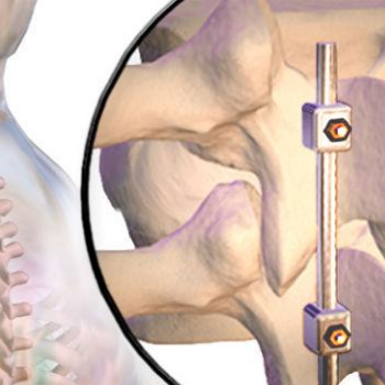
Distraction osteogenesis (DO), also called callus distractioncallotasis and osteodistraction, is a process used in orthopedic surgery, podiatric surgery, and oral and maxillofacial surgery to repair skeletal deformities and in reconstructive surgery. The procedure involves cutting and slowly separating bone, allowing the bone healing process to fill in the gap.

Medical Uses

Distraction osteogenesis (DO) is used in orthopedic surgery, podiatric surgery, and oral and maxillofacial surgery to repair skeletal deformities and in reconstructive surgery. It was originally used to treat problems like unequal leg length, but since the 1980s is most commonly used to treat issues like hemifacial microsomia, micrognathism (chin so small it causes health problems), craniofrontonasal dysplasias, craniosynostosis, as well as airway obstruction in babies caused by glossoptosis (tongue recessed too far back in the mouth) or micrognathism.

In 2016 a systematic review of papers describing bone and soft tissue outcomes of DO procedures on the lower jawbone was published; the authors had planned to do a meta-analysis but found the studies were too poor in quality and too heterogeneous to pool. From what they were able to generalize, the authors found there was significant relapse in the vertical plane for bone, and a higher risk of relapse when there was an initial high gonial angle or Jarabak ratio (sella–gonion/nasion–menton). For soft tissue, little evidence was available regarding the vertical dimension, while a 90% correspondence between skeletal and soft tissue was found for sagittal positioning; the dental-to-soft tissue agreement was around 20%.

A 2016 Cochrane review of DO on the upper jawbone to treat cleft lip and cleft palate compared with orthognathic surgery found only one study, involving 47 participants and performed between 2002 and 2008 at the University of Hong Kong. This was not sufficient evidence from which to generalize, but the authors noted that while both procedures produced notable hard and soft tissue improvements, the DO group had greater advancement of the maxillary and less horizontal relapse five years after surgery. There was no difference in speech or nasal emissions outcomes nor in adverse effects; the DO group had lower satisfaction at three months after surgery but higher at two years.
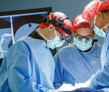
Distraction osteogenesis

Procedure

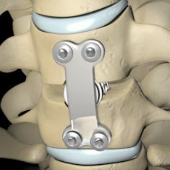
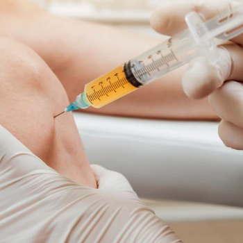
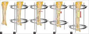
In the first phase, called the “osteotomy/surgical phase”, the bone is cut, either partially, only through the hard exterior, or completely, and a device is fitted which will be used in the next phases. In the second phase, the latency period, which lasts generally seven days, the appliance is not activated and early stages of bone healing are allowed. In the third phase, the “distraction phase”, the device, which is mounted to the bone on each side of the cut, is used to gradually separate the two pieces, allowing new bone to form in the gap. When the desired or possible length is reached, which usually takes three to seven days, a consolidation phase follows in which the device keeps the bone stable to allow the bone to fully heal. After the consolidation phase, the device is removed in a second surgical procedure.

The device that is used is usually manually operated by twisting a rod that through a rack and pinion system or the like, separates the bone; the rate of separation is carefully determined because going too quickly can cause nonunion, in which unstable fibrous connective tissue is formed instead of bone, and going too slowly can allow premature union to occur. Generally the rate is about a millimeter per day, achieved in two steps per day. The frequency of steps and how much the device is moved at each step, is called the “rhythm”. The devices sometimes contain a spring that provides tension to continually separate the bones, instead of being manually operated at set intervals.


Distraction osteogenesis


Despite these manually operated systems there are also motorized systems like the FITBONE from WITTENSTEIN. The FITBONE is a fully implantable, motorized, lengthening and correction nail. Advantages of this device are accurate deformity correction, low scar tissue formation, and reduced risk of infection. Furthermore the patients describe the procedure as more comfortable than limb lengthening with mechanical systems.

Content credit to wikipedia.org

Image credit to researchget.net

Anterior cervical discectomy and fusion(ACDF)

Anterior cervical discectomy and fusion (ACDF) is a surgical procedure to treat nerve root or spinal cord compression by decompressing the spinal cord and nerve roots of the cervical spine with a discectomy, followed by inter-vertebral fusion to stabilize the corresponding vertebrae.This procedure is used when other non-surgical treatments have failed.

Medical uses

ACDF is used to treat serious pain from a nerve root  that has become inflamed. This can be caused by:

1. a herniated disc when other non-surgical treatments have failed. The nucleus pulposus (the jelly-like center of the disc) of the herniated disc bulges out through the annulus (surrounding wall) and presses on the nerve root next to it.

2. degenerative disc disease (spondylosis). The disc consists of about 80% water. When one grows older, the disc starts to dry out and shrink, causing small tears in the annulus and inflammation of the nerve root.


Technique

Anterior cervical discectomy and fusion(ACDF) surgery

The neurosurgeon or orthopedic surgeon enters the space between two discs through a small incision in front (= anterior) of and at the right or left side of the neck. The disc is completely removed, as well as arthritic bone spurs. The disc material, pressing on the spinal nerve or spinal cord, is then completely removed. The intervertebral foramen, the bone channel through which the spinal nerve runs, is then enlarged with a drill giving the nerve more room to exit the spinal canal.

To prevent the vertebrae from collapsing and to increase stability, the open space is often filled with a graft. That can be a bone graft, taken from the pelvis or cadaveric bone; or an artificial implant. The slow process of the bone graft joining the vertebrae together is called “fusion”. Sometimes a titanium plate is screwed on the vertebrae or screws are used between the vertebrae to increase stability during fusion, especially when there is more than one disc involved.

Anterior cervical discectomy and fusion(ACDF)  surgery


Recovery

The surgery requires a short stay in the clinic (1 to 3 days) and a gradual recovery between 1 and 6 weeks. However, the technology has advanced and it can be performed by ‘Endoscopic Micro Discectomy” with the patient able to continue their normal life in two days. The patient may be advised to wear a neck brace or collar (for up to 8 weeks) that serves to ensure proper spinal alignment. Wearing the brace heightens one’s awareness of posture and positioning and helps prevent movements (e.g., sudden and/or excessive bending or twisting of the neck) that may aggravate or slow down the healing process.

It is especially advisable to wear a protective neck brace when traveling (e.g., by car), sleeping, showering, or any other activities in which the patient may not be able to be ensure proper spinal alignment. In addition, physical therapy and related healing modalities (e.g., massage, acupuncture) may be recommended in order to promote proper healing, as well as to strengthen the surrounding muscles that can take over the neck brace’s ‘job’ of ensuring proper spinal alignment when the patient starts (around 4 to 6 weeks after surgery) to wean off the neck brace.

Article From : wikipedia.com

You know what is Minimally Invasive Spine Surgery?

Spine surgery has advanced dramatically with the technology surrounding minimally invasive surgical (MIS) techniques. Surgical materials, nerve monitoring, and computer-aided navigation have improved significantly recently. With these techniques, the risks, recovery time, and surgical complications have decreased greatly.

Minimally Invasive Spine Surgery

For some patients who have spinal instability or have been recommended spinal fusion, it is important to understand what minimally invasive options may be available.

Advances in spine imaging, such as MRI, allow surgeons to see smaller and smaller spinal problems. Individual spinal nerves, bone spurs, and small disc fragments can be easily seen and diagnosed. Similarly, spinal surgery techniques have advanced to the point where some procedures involve incisions less than 1 inch.

Many modern techniques can be described as “minimally invasive” surgery, or “MIS.” For some patients, a minimally invasive procedure may be a better surgical option than a traditional or “open” spinal surgery. Instead of removing muscle attachments from bones, a minimally invasive surgery spreads and pushes muscles out of the way.

X-rays, computer-aided navigation techniques, and specialized tubes or retractors with special cameras or microscopes have led to major advances in the capabilities of MIS surgery. In addition to a smaller incision, MIS surgery has several advantages. It has been shown, on average, to cause decreases in blood loss, post-operative pain and narcotic use, soft tissue damage, and number of days spent in the hospital. On average, patients have a faster recovery, and return to normal activity and work faster.

In general, most spinal surgery can be grouped into 2 categories:

  1. Decompression
  2. Fusion

Procedures to decompress nerves of the spine include removing: herniated discs, enlarged ligaments and soft tissue, and bone spurs that are pressing on spinal nerves. In some cases, motion of the spine may become unstable or painful, either from wear and tear, trauma, or from a decompression procedure. These patients may be offered spinal fusion. There are MIS options and techniques available for both types of spinal surgery.

Minimally Invasive Decompression

A normal spinal canal is filled with fluid that surrounds the spinal cord and nerves. As the spine degenerates, spinal joints get bigger (just like the knuckles on our fingers), and discs may collapse or bulge. As this happens, room for the spinal cord and roots decreases and this may lead to pain or weakness in the legs. In severe cases, a minimally invasive surgical procedure may not be an option, and an “open” procedure may be the best choice to remove arthritic bone spurs. However, for some patients where spinal stenosis is in only 1 or 2 levels, a minimally invasive surgery may be a better option with a quicker recovery.

Bone spurs and arthritis is removed from the spine where nerves are compressed using a tube or retractor that is often less than an inch in diameter. A high-powered surgical microscope is used through the tube so that the nerves can be safely seen and protected throughout the operation. This smaller incision, less muscle disruption, and preservation of more of the patient’s anatomy may lead to a quicker recovery, and physical therapy can begin sooner.

Minimally Invasive Spinal Fusion

Spinal fusion is always a major undertaking, and any patient considering it should understand the risks and recovery involved. Damage to nerves, infection, failure of bones to fuse properly, or the need for more surgery is all included in the risks of a minimally invasive or traditional “open” surgery. For this reason, it is important that patients consider and fail all possible non-surgical or “conservative” methods with their surgeon before making the decision to undergo spine surgery. These non-surgical treatments include physical therapy, heat/ice, anti-inflammatory or neurologic-acting medication, bracing, or spinal injections before surgery.

Instrumentation used to fuse vertebrae together is placed under x-ray guidance through small and specialized retractors, with an incision about an inch and a half or less in length. Through the same retractors, bone spurs are removed, and spinal nerves are decompressed using a high-powered surgical microscope. For these patients, a smaller incision and less muscle disruption may lead to a quicker recovery and less post-operative pain.

The final tube requires an incision that is less than one inch, and minimizes soft tissue damage.

The final tube requires an incision that is less than one inch, and minimizes soft tissue damage. Reprinted with permission © bjerkespine.comWhen spine surgery is recommended, it is important for patients to discuss minimally invasive options with their surgeon.

Minimally invasive surgery, or “MIS,” is not the right option for all patients. It is important to understand why you may not be a candidate for MIS. Some surgeons have not been trained using these techniques, and do not offer them even though their patients may benefit. Before undergoing a spinal surgery, you should have a good understanding of the procedure itself and what to expect during the recovery process. Since pain and recovery are personal and individualized for each patient, no surgeon can guarantee a particular recovery course. However, for many patients, an MIS option may result in a smoother and more successful outcome.

Postoperative Care for Spinal Fusion Surgery

The extended healing period required after lumbar spinal fusion surgery makes postoperative care especially important. While spinal fusion surgery has a high success rate for stabilizing 2 or more adjacent vertebrae and enabling a return to previous normal activity levels, the recovery time can vary based on many factors. These factors include the extent of the surgery, other medical conditions, and how closely the care instructions are followed.

Spinal Fusion Recovery Time

Most people are able to return home from the hospital about 2 to 4 days after lumbar spinal fusion surgery (if there are other people at home). Driving may be resumed a couple weeks after that if off opioid medications. It typically takes about 4 to 6 weeks to return to an office or sedentary job, but it can take 3 months or longer to return to activities that are more physical.

Despite the name of the surgery, the spine is not actually fused during a lumbar spinal fusion procedure. Instead, during the surgery a bone graft or substitute is placed in the spine that facilitates bone growth between the adjacent vertebrae to eventually form one bone, a process that usually takes about 3 to 6 months. The new bone will immobilize the spine at that segment. Screws, cages, plates, and rods may be implanted during surgery to stabilize the area while the bone heals and becomes solid. Some patients also wear a brace during recovery that limits motion.

The bone continues to mature and solidify over 12 to 18 months after the surgery. Many people with a single-level fusion are able to return to all activities even vigorous ones such as weightlifting or construction work—about 6 months after surgery.

Walking and Moving After Spinal Fusion Surgery

Keeping the spine aligned correctly is important after surgery in order to minimize its workload and reduce the risk of disrupting the healing process. Patients work with physical and occupational therapists each day to learn the safest ways to dress, sit, stand, walk, and take part in other activities without putting added stress on the back. Even getting out of bed requires a special technique—known as log-rolling—to avoid twisting the spine.

In some cases, the physical therapist may advise the patient to use a walker for stability. The occupational or physical therapist also helps arrange medical equipment for later use in the person’s home, if needed.

Many people find it helpful to bring sturdy slip-on shoes with them to the hospital, since surgeons and hospital staff encourage patients to get up and walk around as quickly as possible after the surgery.


From : spine-health.com

What are the Most Common Types of Orthopedic Surgery?

An orthopedic surgery is any operation performed on the musculoskeletal system. This system is comprised of your bones, muscles, ligaments, joints and tendons. There are three different types of orthopedic surgery. Traditional procedures are now competing with minimally invasive arthroscopic surgeries that tout less pain and quicker recovery times.

Let’s take a look at the most commonly performed orthopedic procedures.

    • Joint Replacement Procedures. These procedures replace an injured joint with a prosthetic and are among the most common orthopedic operations. Common joint replacement surgeries include hip and knee replacement surgeries. It is important that patients are monitored for signs of complications after these procedures because the procedures carry a considerable amount of risk. Among these risks are the chances that the implant will fail or that the materials making up the implant will make their way into the blood, causing a toxic condition known as metalosis.
    • Revision Joint Surgery. If an existing implant has failed, it may be necessary to remove it and implant a new one. Revision surgeries are often required when the patient received a defective implant or an older implant has failed.
    • Debridement. Whenever tissue death has occurred and the affected tissue needs to be removed before healing can occur, a debridement procedure is how doctors will remove it. There are some cases where bone is also removed when necessary.
    • Spinal Fusion. Spinal fusions join the vertebrae together to provide more stability to the spine or to repair damage to the spine.
    • Bone Fusion. Like spinal fusions, bone fusions use grafting to fuse fractured bones together so that they can heal.
    • Soft tissue repair. These procedures focus on torn ligaments or tendons.
    • Internal Fixation of Bones. This type of surgery places fragments of bones together and keeps them in place using pins, screws or plates so that they can heal. In some cases, the devices will remain inside of the body.
    • Osteotomy. If a child has bone deformities, he or she will need this type of operation to help correct the deformity so that the bone grows properly.

From : www.rosenfeldinjurylawyers.com

Sarcopenia’s Role in Knee OA Progression

The incidence of total knee arthroplasty to treat end-stage knee osteoarthritis (OA) continues to rise even in the face of patient risk-stratification tools and alternative payment models. Consequently, payers, patients, and their doctors are placing a premium on methods to prolong the native knee joint and delay or avoid surgery. This partly explains the explosion of interest in biologics and the subsequent checkreins being put in place regarding their use.

As the AAOS clinical practice guidelines for the management of knee arthritis clearly state, the best management for symptoms of knee arthritis remains weight loss and self-directed physical activity. However, there is uncertainty regarding which subtypes of patients are likely to achieve OA symptom benefits with different weight-loss strategies.

A recent large, multicenter cohort study published in Arthritis & Rheumatology attempted to further characterize patient body composition and its association with knee OA.  Using whole-body dual x-ray absorptiometry (DXA) measures of fat and muscle mass, researchers classified patients into one of four categories: nonobese nonsarcopenic, sarcopenenic nonobese, nonsarcopenic obese, or sarcopenic obese. Sarcopenia is the general loss of muscle mass associated with aging. If orthopaedic surgeons better understand how fat and muscle metabolism change with time and affect inflammation and chronic disease, they may be able to provide patients with additional insight into preventive measures.

Using DXA-derived calculations, the authors observed that among older adults, the relative risk of developing clinically significant knee osteoarthritis (Kellgren-Lawrence grade ≥2) at 5 years was about 2 times greater in both sarcopenic and nonsarcopenic obese male and female patients compared to nonobese, nonsarcopenic patients.  Sarcopenia alone was not associated with risk of knee OA in women or men. In a sensitivity analysis focusing on BMI, men showed a 3-fold greater risk of knee OA if they were sarcopenic and obese, relative to nonobese nonsarcopenic patients.

The takeaway from this study is that focusing solely on fat/weight loss may overlook a valuable opportunity to slow the progression of knee arthritis in some patients.  Further studies are needed to validate the contribution of low muscle mass to the development and progression of symptomatic knee arthritis.

 Credit to : Jeffrey Stambough, MD(royortho.com)

Postoperative Care for Hip Replacement

Before hip replacement surgery even takes place, a surgeon and patient will talk about a recovery and rehabilitation plan. This plan can help the patient:

  • Leave the hospital sooner
  • Regain hip strength and function more quickly
  • Reduce the risk of developing a post-surgical limp
  • Resume independent living sooner

People are typically able to take care of themselves and resume most activities 6 weeks after surgery, and are 90% recovered after 3 months. It can take up to a full year before they are 100% recovered.

Faster and slower recoveries
Healing and rehabilitation times vary among patients. Deviation from the “typical” recovery timeline cannot always be predicted, but differences are usually okay as long as the patient, physical therapist, and doctor continue to work together towards a full recovery.

Waking Up from Hip Replacement Surgery

A patient may wake up from anesthesia with a triangle-shaped pillow between his or her legs, keeping the legs slightly spread. This pillow is meant to stabilize the hips. A doctor may suggest a patient use the pillow while sleeping and resting in bed during the days following surgery.

In the hours after surgery, the patient will begin to regain feeling in his or her legs. The doctor will pre-emptively treat pain using a combination of pain-relief methods that complement each other and minimize side effects, an approach that is called multimodal analgesia.

If pain is reasonably under control, the patient may be asked to do gentle exercises in bed, such as:

  • Bending and flexing the ankle (ankle pump)
  • Contracting and relaxing the quadriceps muscles
  • Contracting and relaxing the gluteal muscles

Gentle exercises may be done for few minutes each hour (interrupting sleep is not necessary) to facilitate blood flow.

The patient may be asked to stand up and take a few steps with the aid of a physical therapist and/or a walker. Patients who get up and bear some weight (with assistance) on their new hips soon after surgery tend to recover more quickly than patients who do not.

How Much Weight Can Be Put On the New Hip?

A surgeon or nurse will give each patient weight-bearing guidelines to follow in the hospital and at home. Initially, a patient may be advised to put just a small percentage of weight on the affected leg, with incrementally more weight being applied over time.

Exactly how much pressure the new hip can bear will depend on factors such as:

  • The type of surgery and prostheses used
  • The condition of the patient’s natural bone
  • How the prostheses was fixated to the natural bone

For example, patients who have a cementless prostheses may need to wait longer to put weight on the hip, because the bone tissue needs time to grow and bond with the prostheses.

Full Credit To : arthritis-health.com

The role of malnutrition in 90-day outcomes after total joint arthroplasty

Via a cohort study of 4,047 cases, the researchers sought to determine the role of preoperative albumin in total joint arthroplasty (TJA) patients for prediction of length of stay (LOS) and 90-day outcomes. For malnutrition, the albumin cutoff of ≤3.5 g/dL is used as proxy, however the value remained understudied. In some patients at risk for adverse events post TJA, the pre-operative albumin level could be missing. For 90-day readmission, the optimal albumin cutoff was concluded as 3.94 g/dL in a univariable model. Screening for malnutrition as a preoperative evaluation could serve a role. The albumin cutoff value of 3.5 g/dL might be missed.

Read the Full Article on :

https://bit.ly/2QlIuY1

Efficiency of platelet-rich plasma therapy in knee osteoarthritis does not depend on level of cartilage damage

Platelet-rich plasma (PRP) is a concentrate of autologous blood growth factors which has been shown to provide some symptomatic relief in early osteoarthritis (OA) of the knee. Platelet-rich plasma therapy is a simple and minimally invasive intervention which is feasible to deliver in primary care to treat osteoarthritis of the knee joint.

Researchers ascertained if patient satisfaction with platelet-rich plasma (PRP) therapy was correlated with the degree of cartilage damage quantified with magnetic resonance imaging (MRI). A pre-treatment MRI was performed and analyzed according to Peterfy and colleagues by a senior consultant radiologist with the whole-organ MRI scoring method (WORMS) to assess the level of osteoarthritis. They performed PRP in 59 patients using a low-leukocyte autologous conditioned plasma system. Findings suggested that intraarticular injection of PRP may improve symptoms of osteoarthritis and decrease pain in patients with knee joint osteoarthritis regardless of the level of cartilage damage quantified by the whole-organ MRI scoring method WORMS.

Credit to : www.mdlinx.com

Orthopedic Implants to Dominate the Global 3D Printed Medical Devices Market

Increasing prevalence of chronic diseases and rising awareness regarding personal care will drive future growth.

The global 3D printed medical devices market is expected to expand at a compound annual growth rate (CAGR) of 18.1 percent through 2027. An increasing prevalence of chronic diseases and rising awareness regarding personal care are major factors twill drive growth of the market during this period, according to Future Market Insights.

Increased Ability to Innovate Aids the Global 3D Printed Medical Devices Market
With increasing popularity of 3D printing, medical devices manufacturers are particularly focused on innovations. Substantial need for individualized yet economical medical solutions is met through 3D printing. Complex features of integration such as hard and soft areas, solid and porous structures, multi-material and multi-color, which seemed to be difficult by implementing conventional manufacturing techniques, is made simpler through 3D printing. Patient-specific implants are being manufactured based on computed tomography (CT) and magnetic resonance imaging (MRI) scans provided by surgeons, which results in reduction of the overall surgical cost. This has increased the popularity of 3D printed medical devices all around the world.

The global 3D printed medical devices market is segmented based on application, technology, material type, end user and region. On the basis of application, the market has been segmented into orthopedic Implants, dental implants and cranio-maxillofacial implants. Orthopedic implants application segment accounted for a higher revenue share in global 3D printing devices as compared to others in application segment. Orthopedic implants segment in the application category of the global 3D printed medical devices market was estimated to be valued at nearly $170 million in 2017 and is slated to reach a valuation of nearly $970 million in 2027, exhibiting a 19.2 percent CAGR during the period of assessment.

Based on the material type, 3D printed medical devices market is segmented into metals and alloys, biomaterial inks and plastics. The biomaterial inks segment was estimated to be valued at nearly $65 million in 2017 and is anticipated to reach a valuation of nearly $400 million in 2027, displaying a CAGR of 20 percent during the period of forecast.

Based on the end user, 3D printed medical devices market is segmented into hospitals, ambulatory surgical centres and diagnostic centres. Hospital end user segment accounts for higher demand for 3D printing devices as compared to other distribution channel segments such as ambulatory surgical centres and diagnostic centres, registering a CAGR of 18.7 percent over the forecast period.

On the basis of region, the global 3D printed medical devices market has been segmented into North America, Latin America, Western Europe, Eastern Europe, Asia Pacific excluding Japan, Japan and the Middle East and Africa. Revenues in Western Europe are expected to grow at a CAGR of 18.6 percent, whereas Eastern Europe is anticipated to grow at 15.5 percent over the forecast period. The Western Europe 3D printed medical devices market was estimated to be valued at nearly $65 million in 2017 and is slated to reach a value of nearly $360 million in 2027. The Eastern Europe 3D printed medical devices market was estimated to be valued at nearly $40 million in 2017 and is anticipated to reach a value of nearly $160 millin in 2027.

Some of the players in the global 3D printed medical devices market include 3D Systems Inc., Arcam AB, Stratasys Ltd., FabRx Ltd., EOS GmbH Electro Optical Systems, EnvisionTEC, Cyfuse Biomedical K.K. and Formlabs Inc., among others. Key players are focusing on strengthening their position by establishing new facilities in North America region. Moreover, companies are targeting Asia-Pacific region and Europe by distribution agreements with local players. In order to increase their revenue, major players in the North America region are entering into agreements with hospitals and academic institutes.

Future Market Insights provides market intelligence and consulting services, serving clients in over 150 countries. FMI is headquartered in London and has delivery centers in the United States and India.

From : www.odtmag.com

    We offer a wide variety of safe and advanced Orthopedic Implants and fixators. At the helm of the company’s operations is Mr. Vinodbhai, the Director. His enterprising skills and experience in the industry continue to play a pivotal role in the company’s growth.We are based in Ahmedabad city West Part of India.

    ADDRESS

    Plot No.10, Phase-1, B|h. Prashant Eng., G.I.D.C.Vatva, Ahmedabad-382 445, Gujarat, (INDIA).

    PHONE

    +91 9375801932

    EMAIL

    [email protected]

    Cart